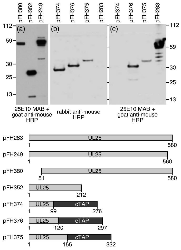
Fig. 1.

SDS–PAGE and immunoblotting of truncated UL25 proteins or UL25-TAP fusion proteins. Plasmids expressing different UL25 proteins were transfected into Vero cells. Proteins from cell lysates were isolated 48 h post-transfection separated by SDS–PAGE, and then electrophoretically transferred to a nitrocellulose membrane and blotted (a and c) with UL25E10 mAb followed by a goat anti-mouse HRP conjugated antibody or (b) with a rabbit anti-mouse HRP conjugated antibody. Molecular mass standards are shown (kDa) to the left and right.
