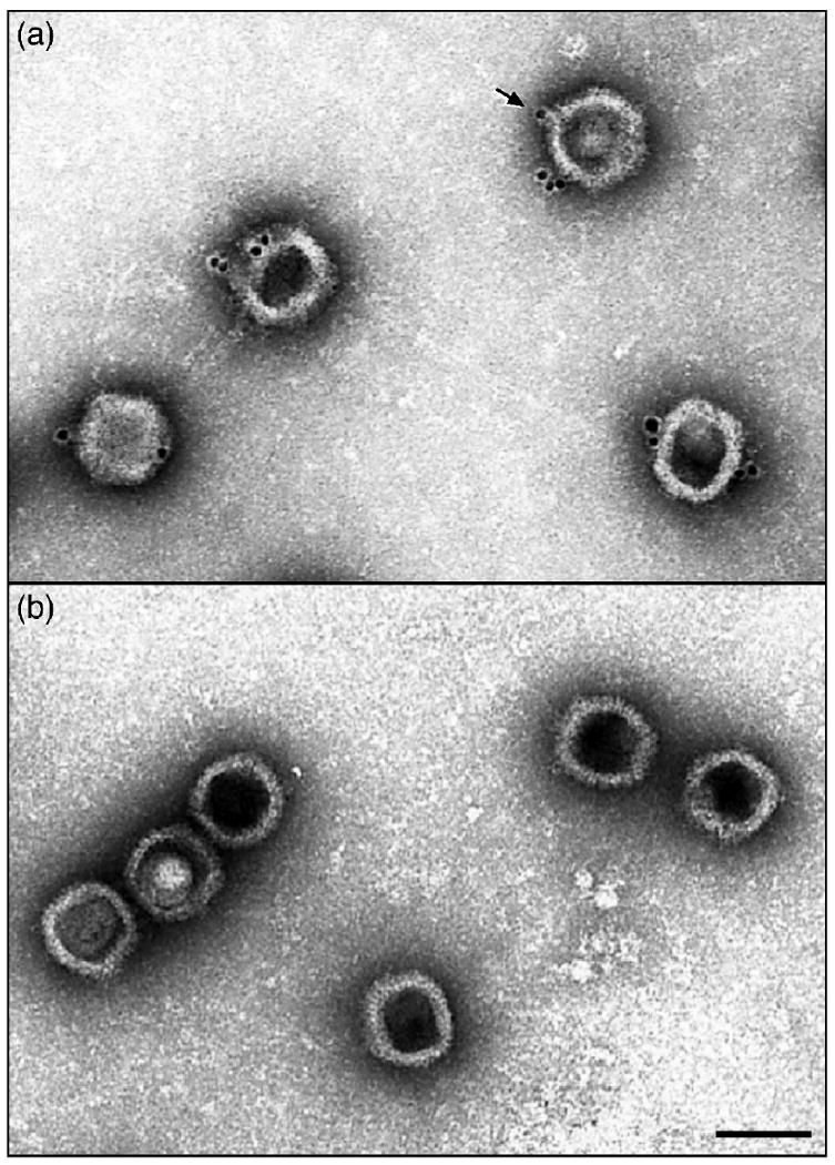
Fig. 3.

Electron micrographs showing (a) C capsids isolated from KOS infected cells or (b) B capsids isolated from UL25 null virus infected cells after staining with the UL25 mAb 25E10 followed by an anti-antibody conjugated to gold beads. Note that the gold beads are found only on the C capsids and appear to be located at the capsid vertices (one is indicated by an arrow) and the majority of the C capsids have labels at more than one site. Bar = 1000Å.
