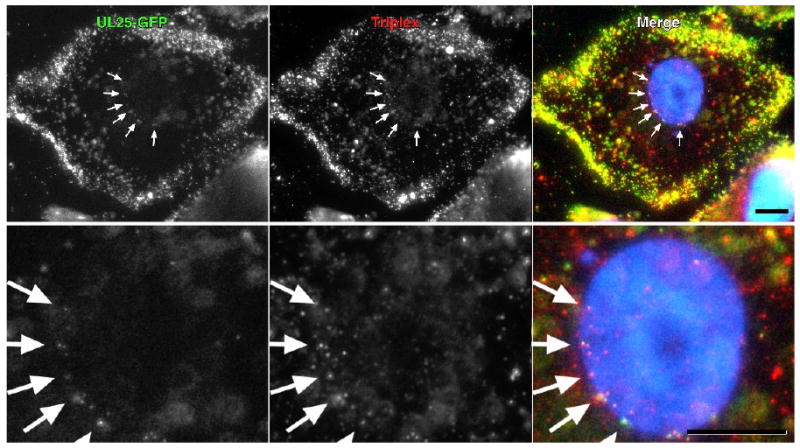
Fig. 6.

Colocalization of UL25-GFP and capsid triplex protein VP23 by fluorescence microscopy. Vero cells were infected for 3 h with vUL25-GFP and the colocalization of the capsid associated UL25-GFP with the capsid triplex protein VP23 was examined in cells that were fixed and immunostained for VP23 (red). Nuclei were DAPI stained. Note that the UL25-GFP and VP23 protein labels occur coincidently in the cytoplasm and at the nuclear surface (arrows). Bar = 10 um. A close-up image of the nuclei for each of the upper panels is shown in the lower panel.
