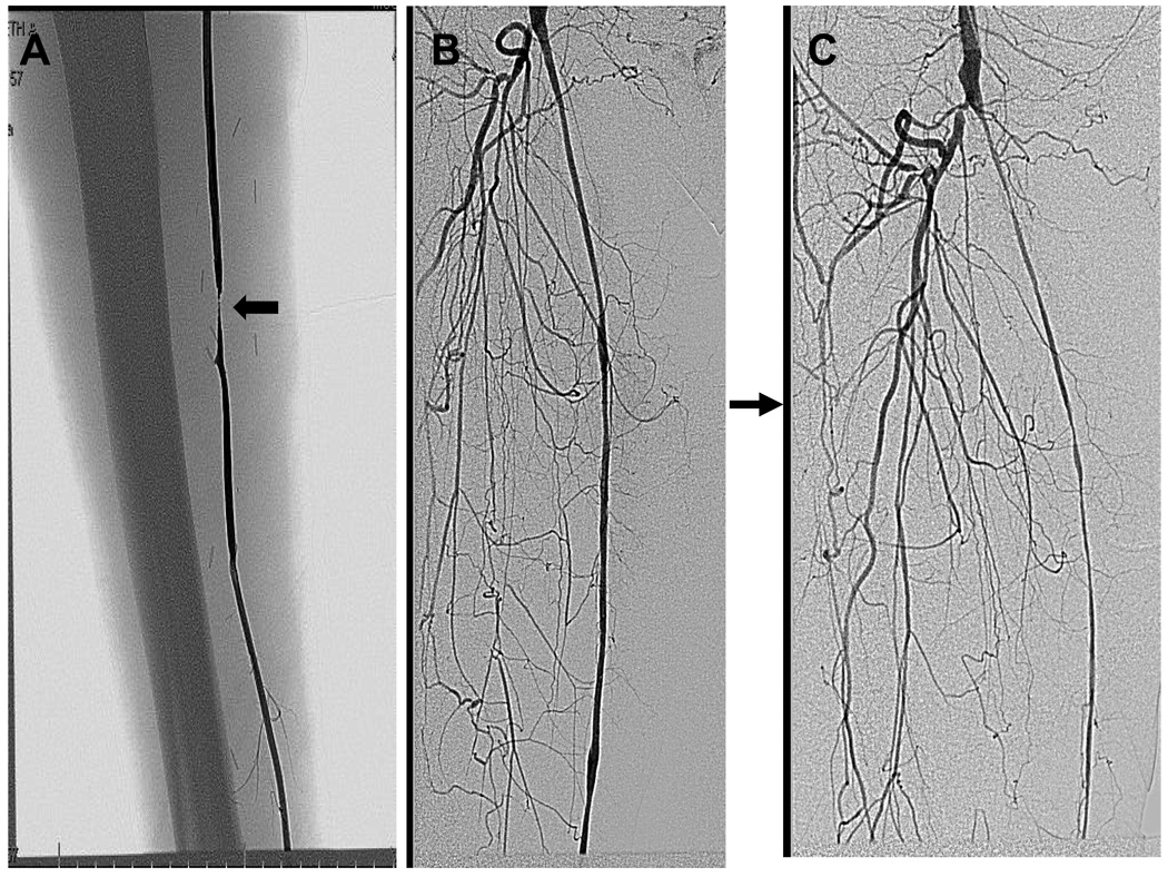
Figure 3.
Modes of vein graft failure. While the majority of intimal hyperplasia in vein grafts is focal, a substantial minority (12%) is diffuse. Panel A represents a 6 month old vein graft in a 67 year old white man that developed a mid graft stenosis (arrow) that was successfully treated with a vein patch angioplasty. Panel B represents a 4 month old vein graft in a 77 year Black women undergoing angiography for contralateral limb ischemia. Three months later, she developed diffuse intimal hyperplasia which progressed to vein graft occlusion.

