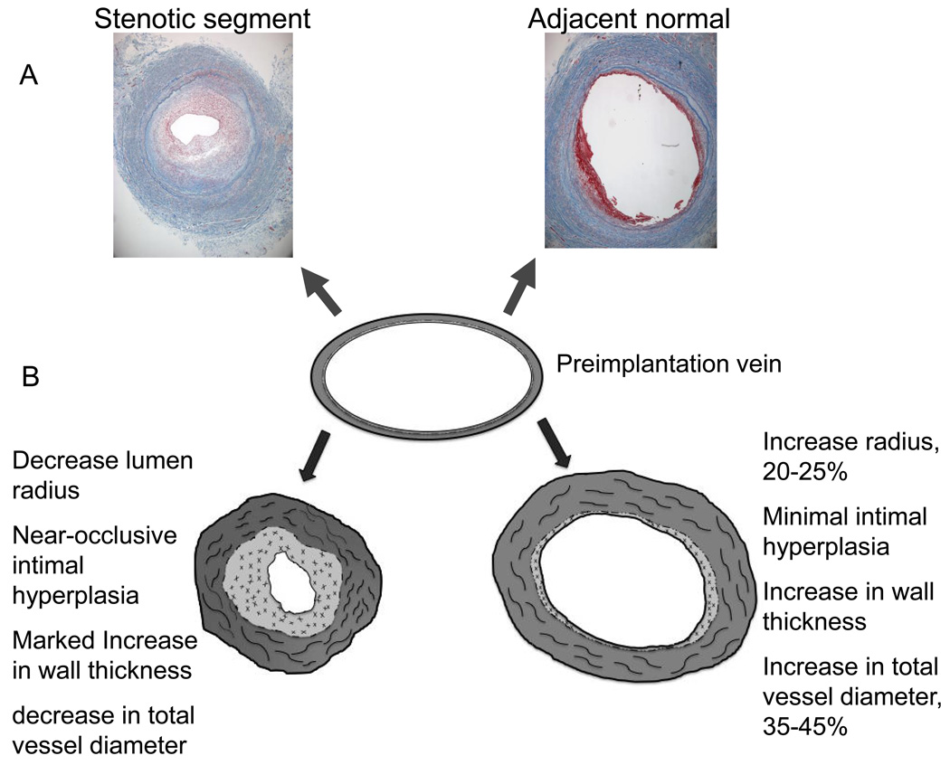
Figure 4.
Histologic sections (10X) of an 8 month old vein graft which developed a focal mid-graft stenosis and underwent open revision with a short interposition graft. The vein was of uniform size and caliber at the time of implantation. The sections were taken approximately 2 cm from one another. The area of stenosis has developed marked intimal hyperplasia and has a smaller area circumscribed by the internal elastic laminae as well as decreased total vessel diameter indicative of negative remodeling of the entire vein graft, A. Theoretical normal and abnormal adaptation patterns of a human lower extremity vein grafts, B. Normally the lumen and wall area increase in the early post-implantation period to produce a lumen diameter to wall thickness ratio of about 7.

