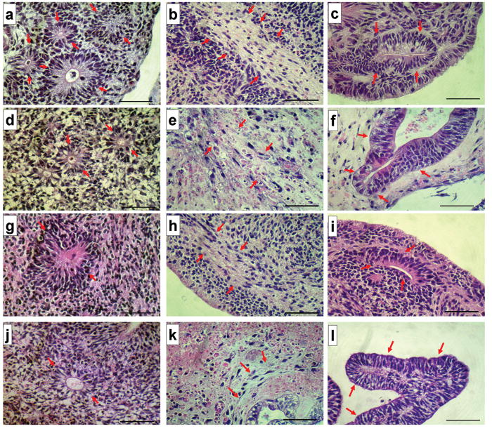
Figure 3.
Histological evidence of Tri-Lineage Differentiation in embryoid bodies generated from hPSCs. Shown are images of hematoxylin and eosin-stained histologic sections of EBs from hESC cells propagated on MEFs (top row, a-c), hiPSC propagated on MEFs (second row, d-f) as positive controls, hESC cells propagated in the MBIC system (third row, g-i) and hiPSC propagated in the MBIC system (bottom row, j-l). Tri-lineage potential is demonstrated as ectodermal (neuroepithelial) differentiation (a, d, g and j); mesodermal (fibrous connective) differentiation (b, e, h and k) and endodermal (intestinal) differentiation (c, f, i and l). Arrows point to the corresponding tissue in each figure. Magnification is 400x total (10x ocular, 40x objective). Each scale bar represents 50μm in length.

