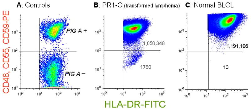
FIGURE 1. GPI (-) populations arise spontaneously in vitro in malignant cell lines.

Flow cytometry pseudo-density dot plots. (A) PIG-A (+) cells and PIG-A (-) cells within a single BLCL from a patient with PNH. The PIG-A (-) population does not express GPI-linked proteins but does express the transmembrane protein HLA-DR. (B) PR1-C, a transformed follicular lymphoma cell line, analyzed after expansion after sorting to eliminate pre-existing mutants. The normal population expresses GPI-linked proteins and transmembrane proteins, registering in the upper right quadrant. There is a large population of spontaneously arising mutants registering in the lower right quadrant that appear similar to the PIG-A (-) cells from the patient with PNH. The mutant frequency, f, is calculated as the number of events in the lower right quadrant divided by the total number events analyzed (see table). (C) A BLCL from a normal donor, demonstrating a much smaller number of spontaneously arising GPI (-) cells.
