
FIGURE 2. Parallel analysis of cultures of malignant cell lines using antibodies specific for transmembrane proteins or the FLAER reagent.
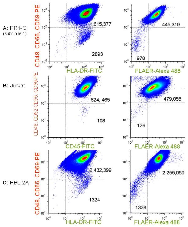
Samples were analyzed after expansion in vitro after sorting to eliminate preexisting mutants. Pseudo-density dot plots are shown for analyses using FITC-conjugated antibodies specific for transmembrane proteins (left panels) or the FLAER Alexa-488 reagent, which binds to the GPI structure directly (right panels). In all 3 examples, there are distinct populations of spontaneously arising cells that neither express GPI-linked proteins nor take up the FLAER reagent, but which do express transmembrane proteins. (A) A subclone of PR1-C (transformed lymphoma); (B) Jurkat, a T cell ALL cell line; (C) HBL2-A, derived from a Mantle Cell Lymphoma.