
Fig. 5.
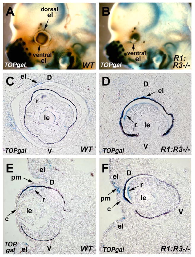
Activation of the Wnt reporter TOPgal in wild-type and RA deficient mouse eyes. (A-B) Staining of whole-mount heads of E12.5 wild-type (WT) and double null (Raldh1-/-;Raldh3-/-) embryos carrying the TOPgal reporter. Arrows indicate TOPgal expression in both the dorsal and ventral eyelid folds of WT, but only in the ventral eyelid fold of the mutant. (C-D) Sagittal sections through the eyes of TOPgal stained E12.5 wild-type (WT) and double null (Raldh1-/-;Raldh3-/-) embryos showing the loss of the normal eyelid folds in the mutant, and ectopic TOPgal expression in the retina. (E-F) Frontal sections showing that the mutant lacks the dorsal eyelid domain, but retains the ventral eyelid domain, and also showing that the mutant has increased TOPgal expression in a central domain of perioptic mesenchyme located where the cornea would normally develop and nearby in the retina. c, cornea; d, dorsal retina; el, eyelid fold; le, lens; pm, perioptic mesenchyme; r, retina; v, ventral retina.