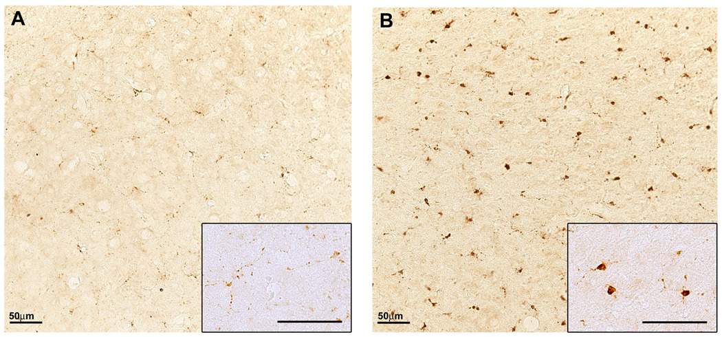
Figure 2. Increased microglial activation in the aged brain.
Immunostaining for the endosomal/lysosomal enzyme CD68, a marker of the microglia/macrophage lineage, shows a prominent increase in the neocortex of 24-month-old C57BL/6 mouse (B) compared with a 6-month-old mouse (A) consistent with increased microglial activation in aged brains. Inserts show individual microglia under higher magnification where hypertrophied cell bodies are evident in aged brains (compare insert A to B). Scale bars represent 50µm under 20 or 40× magnification.

