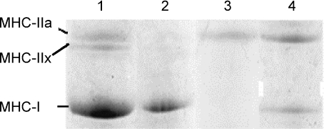
Figure 1. Example of electrophoretic (SDS-PAGE) separation of myosin heavy chain (MHC) isoforms in bioptic samples of isolated fibres from 14-HU rats.
All samples were pure myosin extracted from single muscle fibre. Gels were Coomassie stained. Lane 1 shows a mixed rat fibre sample used as a reference; lane 2, pure slow type I fibre; lane 3, pure fast type IIa fibre; lane 4, hybrid type I–IIa fibre. No type IIx fibres were detected in our experiments.

