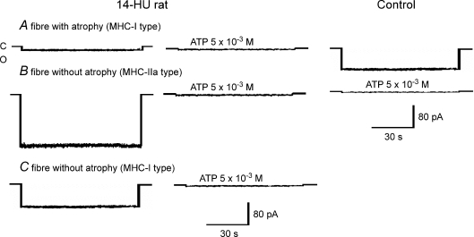
Figure 3. KATP channel currents of SOL muscle fibres from control and 14-day-hindlimb-unloaded (14-HU) rats.
Sample traces of KATP channel currents recorded in excised macropatches from SOL fibres of 14-HU rats and controls during voltage steps from 0 mV holding potential to −60 mV (Vm) with 150 mm KCl on both sides of the membrane, at 20°C. C indicates closed channel levels; O indicates open channel levels. ATP applied on the internal side of the patches inhibited all types of currents. Three types of currents are represented from SOL fibres of 14-HU rats: the first is a sample trace of an atrophic fibre with diameter of 45 μm and KATP current amplitude of > −20 pA characterizing the fibre group named A; the second was not atrophic showing a diameter of 80 μm and a KATP current amplitude of −150 pA characterizing the fibre group named B; the third was not atrophic showing a KATP current amplitude of −85 pA and diameter of 76 μm characterizing the fibre group named C. The KATP current of a control fibre had an amplitude of −84 pA and fibre diameter of 76 μm.

