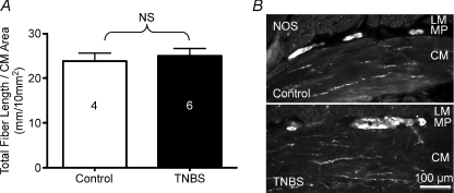
Figure 9. The density of nerve fibres in the circular muscle layer is comparable in the normal colon as compared to the ulcerated region of the TNBS-inflamed colon.
A, graph illustrating measurements of nerve fibres immunostained with antisera directed against NOS and choline acetyltransferase. Immunostaining for both antigens were observed together in circular muscle fibre bundles. B, micrographs of NOS immunoreactivity in sections of normal and inflamed distal colon.

