Figure 4.
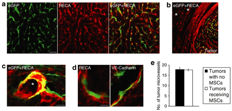
Grafted eGFP+ MSCs closely associate to tumor endothelium and do not express endothelial cell markers. (a) eGFP+ MSCs (green) 8 days following grafting into established N32wt brain tumor. Tumor endothelium is delineated by rat endothelial cell antigen (RECA; red). The majority of mesenchymal stroma cells (MSCs) are closely associated to tumor endothelium. (b) Intratumorally grafted MSCs migrate along RECA+ tumor vessels and do not associate with RECA+ blood vessels in normal brain tissue adjacent to tumor. Asterisks indicate major blood vessels in normal brain. (c) Grafted eGFP+ MSCs closely attached to a major RECA+ tumor vessel. Asterisk indicates tumor vessel lumen. (d) Confocal microscopy analysis was used to determine coexpression of grafted eGFP+ MSCs with endothelial markers RECA or VE-Cadherin within tumors. Grafted eGFP+ MSCs attached to tumor endothelium (RECA and VE-Cadherin, red) and without coexpression of RECA or VE-Cadherin. Bar = 60 µm in a, 100 µm in b, 20 µm in c, and 10 µm in d. (e) Quantification of RECA+ tumor microvessels revealed no difference in tumor microvessel density in tumors receiving eGFP+ MSCs compared to tumors with no MSCs. Data are shown as mean ± SEM, n = 4 in each group. eGFP, enhanced green fluorescent protein; VE-Cadherin, vascular endothelial-Cadherin.

