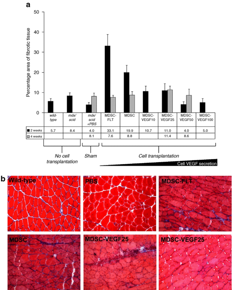
Figure 6.
Fibrosis at site of cellular transplantation. (a) Transplantation of MDSC, MDSC-FLT, MDSC-VEGF10, or MDSC-VEGF25 led to an increase in the amount of fibrosis as measured via trichrome staining, and as compared to noninjected tissue. The greatest amount of fibrosis occurred 2 weeks after MDSC-FLT transplantation, though fibrosis related to transplantation of these cells decreased at 4 weeks post-transplantation. Tissue that was transplanted with cells expressing VEGF (MDSC-VEGF10, MDSC-VEGF25, MDSC-VEGF50, and MDSC-VEGF100; N = 6–16) showed less fibrosis at 2 weeks as compared to tissue transplanted with MDSC alone. By 4 weeks transplantation, there was no significant difference in the amount of fibrosis among the groups (one-way ANOVA). (b) Representative histological sections (×200, Bar = 125 µm). MDSC, muscle-derived stem cell; VEGF, vascular endothelial growth factor.

