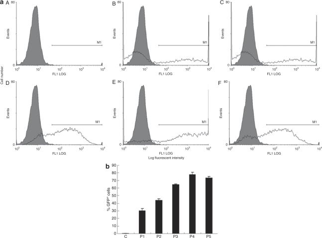
Figure 2.
Flow cytometric analysis of transfection efficacy. (a) Human adipose tissue–derived stem cells were transfected with plasmid pEGFP-N1 and enhanced green fluorescent protein (EGFP)-expressing cells were detected 24 hours later by flow cytometry. Cells were microporated using different voltage and pulse conditions: A, control; B, program 1; C, program 2; D, program 3; E, program 4; and F, program 5. Microporated (white) and control groups (gray) are illustrated in each panel. (b), Comparison of the fraction of GFP+ cells estimated following fluorescence-activated cell-sorting analysis.

