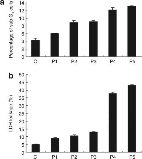
Figure 3.
Analysis of cytotoxic effects of microporation of human adipose tissue–derived stem cells. (a) Flow cytometric analysis. Cells were microporated using different voltage and pulse conditions. After 24 hours, cells were fixed and stained with propidium iodide, followed by fluorescence-activated cell-sorting analysis of DNA content. Sub-G1 populations were defined as the portion of cells with DNA content <2N. The experiment was repeated three times independently. The results are expressed as the mean ± SD. (b) Cells were microporated using different voltage and pulse conditions. After 24 hours, lactate dehydrogenase (LDH) activities were detected as described in Materials and Methods. Results are derived from at least three separate experiments and expressed as the mean ± SD.

