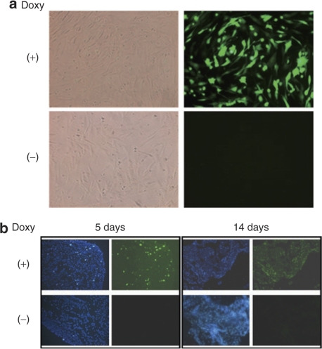
Figure 7.
Microporation of plasmids encoding the Tet-ON system into human adipose tissue–derived stem cells (hADSCs) and testing of enhanced green fluorescent protein (EGFP) expression in vitro and in vivo in response to Dox treatment. First, hADSCs were microporated with pCMV-rtTA and pBI-EGFP plasmids. (a) Following microporation, cells were cultured in the presence or absence of Dox for 24 hours. A strong fluorescent signal was observed in the microporated hADSCs cultured in the presence of Dox (right panel, upper row). In contrast, EGFP expression could not be detected in the Dox (−) group (right panel, lower row). The corresponding phase contrast images are shown in the left panels. Images were taken using ×200 objectives. (b) One day after co-transfection of the pCMV-rtTA and pBI-EGFP plasmids, hADSCs were transplanted subcutaneously on the back of nude mice. The mice were then exposed to drinking water that either contained Dox (200 µg/ml) or not. Cells were harvested at the indicated times. After preparation of frozen sections, EGFP expression was detected by fluorescence microscopy. Images were taken using ×40 objectives.

