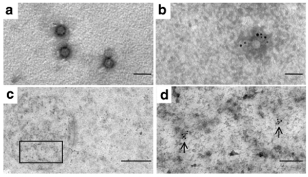
Figure 2.
Identification of adeno-associated viral (AAV)4 particles in the treated retina of P1 by immunogold labeling. (a) AAV4 particles of the injected vector suspension observed by direct electron microscopy. (b) AAV4 particles of the vector suspension detected by immunogold labeling using mouse monoclonal anti-AAV4 antibody ADK4 and goat antimouse antibodies conjugated to 6 nm gold particles. Bar = 50 nm. (c) Epon section of P1 showing the OPL area. The square indicates the area highlighted in d. Bar = 500 nm. (d) Magnification of the area indicated in c. Vector particles are marked with the conjugated gold particles similar to b. Bar = 100 nm.

