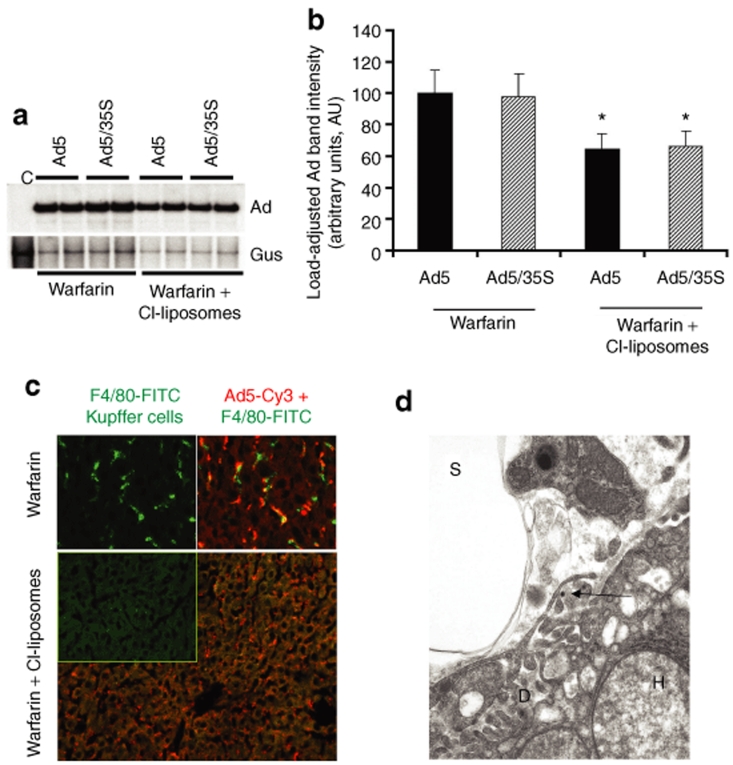
Figure 4.
The treatment of mice with warfarin and clodronate liposomes allows for a partial reduction of the levels of adenovirus (Ad) DNA sequestered by the liver after intravenous virus administration. (a) Southern blot analysis for Ad vector genomes associated with livers of wild-type mice 1 hour after intravenous virus injection. Mice were treated with warfarin only or with a combination of warfarin and clodronate liposomes. Duplicate samples for each group are shown. Gus—mouse β-glucuronidase gene. Control—liver DNA from a mouse injected with virus dilution buffer (phosphate-buffered saline) only. (b) Quantitative representation of Ad accumulation in livers determined by PhosphorImager analysis of Ad-specific bands shown in a after adjustment of Ad DNA signal intensities for the Gus gene signal intensities for corresponding vectors. *P < 0.05. (c) Distribution of Ad particles in the livers of mice treated with warfarin or with a combination of warfarin and clodronate liposomes 1 hour after intravenous virus injection. Liver sections were stained with anti-F4/80 antibody to detect Kupffer cells (green) and with anti-Ad hexon antibody to detect Ad particles (red). Note that the large number of Ad particles was colocalized with Kupffer cells in warfarin-treated animals, whereas Ad particles were associated with liver sinusoids in mice treated with both of drugs. (d) Ad particles (shown by the arrow) localize to a subendothelial Disse space in the livers of mice treated with both warfarin and clodronate liposomes. Electron microscopy analysis was done on ultrathin sections of livers harvested 1 hour after intravenous virus injection. D—Disse space; H—hepatocyte; S—liver sinusoidal space. Magnification ×21,000.

