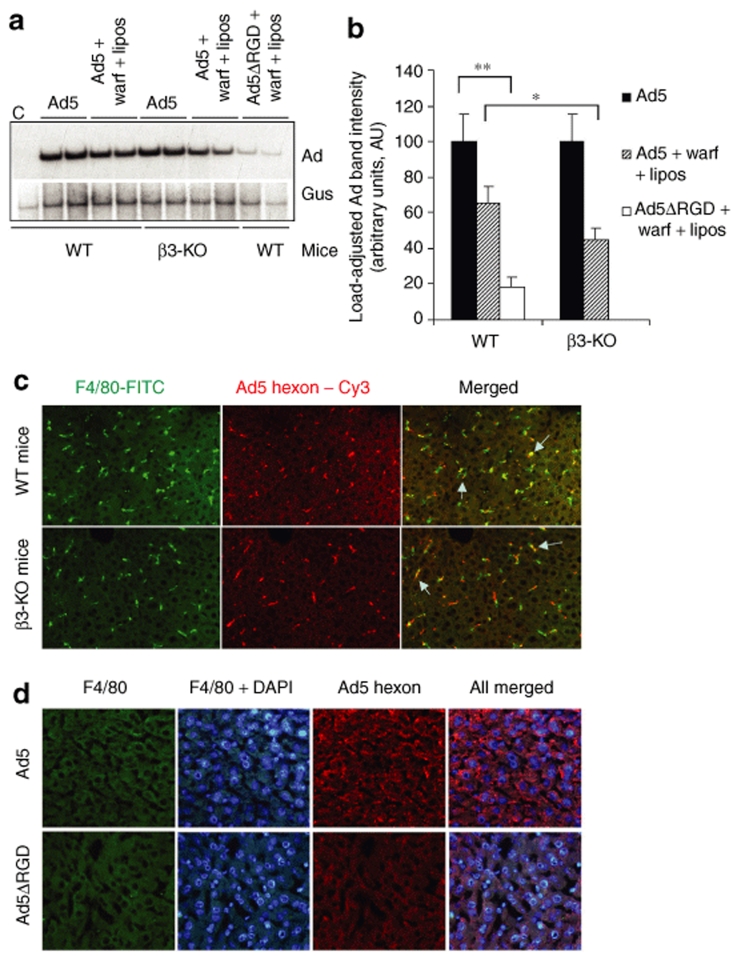
Figure 6.
Adenovirus (Ad) penton RGD motifs play a critical role in supporting the sequestration of the blood-born Ad in the liver. (a) Wild-type (WT) or β3-integrin knockout mice (β3-KO) were treated with a combination of warfarin and liposomes and injected intravenously with Ad5 vector. In addition to drug treatment, WT mice were also injected with Ad5ΔRGD vector. One hour after virus injection, livers were harvested, and total liver DNA was purified and then subjected to a Southern blot analysis as described in Figure 1. Duplicate samples for each group are shown. Gus—mouse β-glucuronidase gene. C—Control liver DNA from a mouse injected with virus dilution buffer (phosphate-buffered saline) only. (b) Quantitative representation of Ad accumulation in livers determined by PhosphorImager analysis of Ad-specific bands shown in a after adjustment of Ad DNA signal intensities for the Gus gene signal intensities for corresponding vectors. *P < 0.05. **P < 0.01. (c) Distribution of Ad particles in the livers of wild-type or β3-KO mice 1 hour after intravenous virus injection. Liver sections were stained with anti-F4/80 antibody to detect Kupffer cells (green) and with anti-Ad hexon antibody to detect Ad particles (red). Colocalization of Ad-specific staining with Kupffer cell staining appears in yellow (merged) and is indicated by arrows. (d) Distribution of Ad and Ad5ΔRGD particles in the livers of mice treated with a combination of warfarin and clodronate liposomes 1 hour after intravenous injection. Liver sections were stained with anti-F4/80 antibody to ensure complete elimination of Kupffer cells with clodronate liposomes (green) as well as with 4′,6-diamidino-2-phenylindole (DAPI) (blue) and anti-Ad hexon antibody to detect Ad particles (red).

