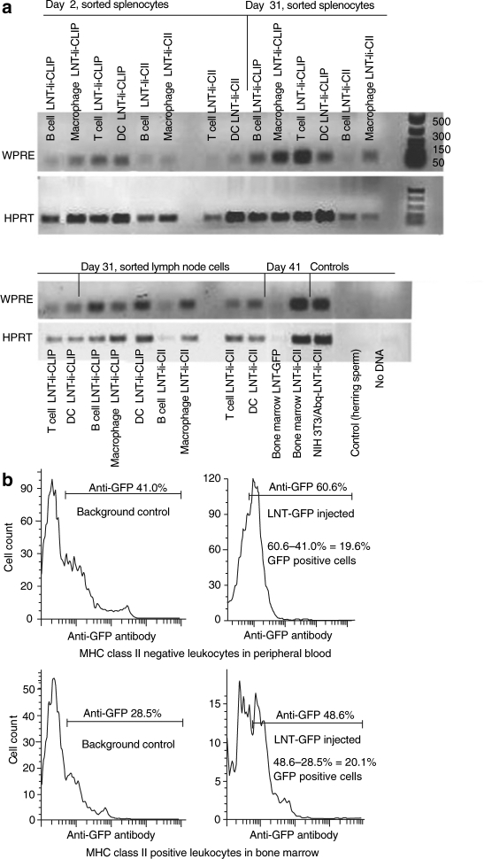
Figure 2.
In vivo detection of the lentiviral vectors. (a) PCR analysis of the woodchuck post-transcriptional regulatory element (WPRE) element in the lentiviral vector in cells sorted from the spleen and lymph node [B cells, T cells, macrophages, dendritic cells (DC)] and in bone marrow cells (not sorted) from mice injected with LNT-Ii-CLIP, LNT-Ii-CII, and LNT-GFP at the indicated times after CII immunization. Aap/Abq+ NIH/3T3 cells stably transfected with LNT-Ii-CII were used as positive controls, and herring sperm DNA (Invitrogen, Sweden), and no DNA were used as negative controls. (b) Flow cytometry analysis of GFP expression in leukocytes in peripheral blood (gated on MHC class II negative cells) and bone marrow (gated on MHC class II positive cells) from control mice (injected with nonvirus) and mice injected with LNT-GFP viral particles 28 days after injection.

