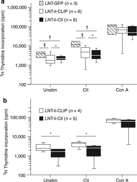
Figure 7.
Proliferation of splenocytes from lentivirus-treated mice and from mice that received bone marrow cells from lentivirus-treated mice. Proliferation of splenocytes taken (a) 40 days after CII immunization from mice injected with LNT-GFP, LNT-Ii-CLIP, and LNT-Ii-CII viral particles and (b) 63 days after CII immunization from mice that received bone marrow from mice injected with LNT-Ii-CLIP and LNT-Ii-CII viral particles. Splenocytes were cultured without any stimuli or in the presence of denatured CII (50 µg/ml) or, as a positive control, Con A (1.25 µg/ml) for 72 hours. Box plots represent median and 75th and 95th centile. Statistical analyses were performed using Student's t-test. *P < 0.05 LNT-Ii-CII versus LNT-Ii-CLIP; †P < 0.05 LNT-Ii-CII versus LNT-GFP; ‡P < 0.05 LNT-Ii-CLIP versus LNT-GFP.

