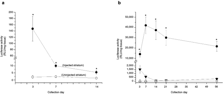
Figure 3.
Luciferase activity in the striatum at various postinjection time points as determined by chemiluminescent analysis. All compacted plasmids used the acetate formulation of CK30PEG10k to form the nanoparticle. (a) Compacted pKCPIRlucBGH plasmid nanoparticles encoding for luciferase and containing the cytomegalovirus promoter was injected into the left striatum only (4.33 µg/µl; 4.0 µl); the right striatum served as a control (uninjected); n = 5 for each treatment. Tissue samples were collected at 3, 7, or 14 days postinjection. Filled circles represent mean luciferase activity (±SEM) on the injected side and measured in relative light units (RLU)/mg tissue while open circles represent mean luciferase activity (±SEM) on the uninjected side; *P < 0.05, compacted versus uninjected side. (b) The pUL3 plasmid encoded for luciferase and contains the polyubiquitin C promoter and pKCIRBGHemptyempty is a control plasmid lacking any promoter or transgene. Nanoparticles were injected into the left striatum only; naked pUL3 (filled triangles; 4.2 µg/µl, 4.0 µl), compacted pUL3 (filled circles; 4.1 µg/µl, 4.0 µl), and compacted pKCIRBGHemptyempty (open triangles; 3.9 µg/µl, 4.0 µl). The right striatum served as a control (open circles; no injection). Symbols indicate mean luciferase activity (±SEM) for each treatment group at each time point. *P < 0.05, compacted versus naked pUL3, compacted pKCIRBGHemptyempty, or no injection; n = 5 for each treatment group.

