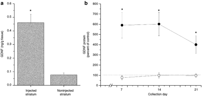
Figure 8.
Glial cell line–derived neurotrophic factor (GDNF) protein expression in the striatum 1–3 weeks following a dual injection of compacted pGDNF into the left striatum. (a) Animals were euthanized 1 week following the injection and both the left and right striata were dissected from the brain. Tissue was analyzed using enzyme-linked immunosorbent assay (ELISA). Bars represent the average (+SEM) GDNF protein values for the injected (left striatum) and the noninjected (right striatum). *P < 0.001, injected striatum versus noninjected striatum. (b) GDNF protein expression in the striatum 1–3 weeks following a dual injection of compacted or naked pGDNF into the left striatum. Animals were euthanized at 1, 2, or 3 weeks following the injection and both the left and right striata were dissected from the brain. Tissue was analyzed using ELISA. Circles represent the average value of GDNF protein (+SEM) calculated as a percentage of the control side (left/right) for compacted pGDNF (filled circles) or naked pGDNF (open circles). Dotted line indicates control levels of GDNF. *P < 0.001, compacted pGDNF versus naked pGDNF.

