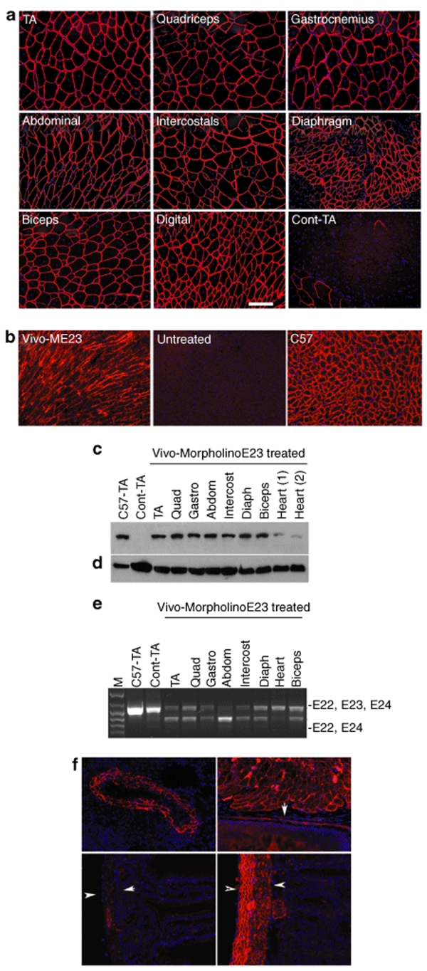
Figure 4.
Restoration of dystrophin expression after five times intravenous injection of 6 mg/kg Vivo-ME23. (a) skeletal and (b) cardiac muscles. (a) TA, tibialis anterior; Digital, right common extensor muscle of forelimb; Cont-TA, TA muscle from untreated mdx mouse. (b) Vivo-ME23, heart from Vivo-MorpholinoE23-treated mdx mouse; Untreated, heart from untreated mdx mouse; C57, heart from normal C57BL/6 mouse. Blue nuclear staining with 4'-6-Diamidino-2-phenylindole (DAPI). Bar = 100 µm. (c) Western blot shows ~50% and >10% of the normal levels of dystrophin in all skeletal muscles and cardiac muscles, respectively. C57-TA, TA muscle from normal C57BL/6 mouse (50% protein loading compared to the other samples); Cont-TA, TA muscle from untreated mdx mouse; Quad, quadriceps; Gastro, gastrocnemius; Abdom, abdominal muscle; intercost, intercostals; Diaph, diaphragm. (d) α-actin as loading controls. (e) Detection of exon 23 skipping by reverse transcriptase-PCR. Total RNA (100 ng from each sample) was used for amplification of dystrophin mRNA from exon 20 to exon 26. Lane M, size marker; Lanes represent the same muscle tissues as described in c. The upper 1,093-bp bands (indicated by E22, E23, E24) correspond to the normal dystrophin mRNA and the lower 880-bp bands (indicated by E22, E24) correspond to the mRNA with exon 23 skipped. (f) Restoration of dystrophin in the smooth muscles of an artery in the lung (top left), muscles of the esophagus (top right, arrow indicate the basal layer of the epithelium of the esophagus) and smooth muscles (layers between the two arrowheads) in small intestine of untreated mdx mouse (bottom left) and Vivo-ME23-treated mdx mouse (bottom right). Blue nuclear staining with DAPI.

