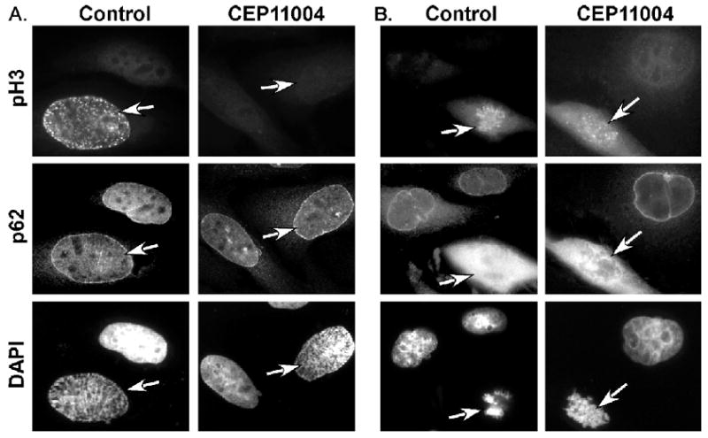
Fig. 8.

CEP-11004 inhibits histone H3 phosphorylation prior to nuclear envelope breakdown. HeLa cells were prepared for immunostaining as described in Fig. 5. Cells were stained for pH3 (top panels) and nucleoporin (p62, middle panels) as a marker of the nuclear envelope. Cells in prophase (A; pre nuclear envelope breakdown) or pro-metaphase (B; post nuclear envelope breakdown) that had been treated in the absence or presence of CEP-11004 were identified. Mitotic cells, as indicated by the arrows, were identified by DAPI staining (bottom panels) of condensed chromosomes. Adjacent interphase cells are shown for comparison.
