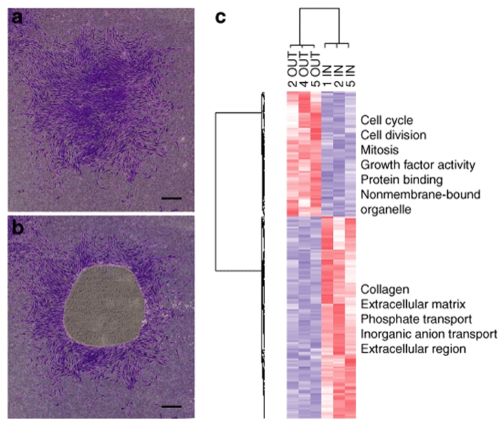
Figure 2.
Demonstration of an in vitro niche found in single cell–generated colony of human MSCs (hMSCs). hMSCs were plated on slides for laser microdissection at 2 cells/cm2 and incubated for 12 days without medium change. The colonies in the figure were stained with crystal violet for illustrative purposes only. (a) Intact colony. (b) Colony after IN region was captured by laser microdissection. (c) Heat map and gene ontologies from microarray data from IN and OUT samples from four colonies (numbers 1, 2, 4, and 5) using 199 differentially expressed genes. Bar in a, and b = 500 µm.

