Figure 1.
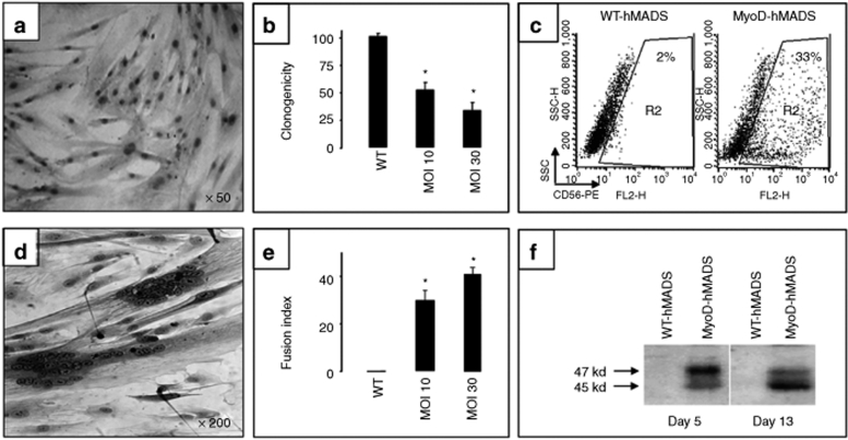
Characterization of MyoD-hMADS cells. (a–c) Transduction of hMADS cells with the lentiviral vector HIV PGK-MyoD. (a) hMADS cells were labeled with an anti-MyoD antibody revealed with peroxydase staining 2 days after transduction. Note the positive nuclear staining of almost all cells. (b) Effect of MyoD on hMADS clonogenicity expressed as the percentage of clones compared to that obtained with WT-hMADS cells (WT). MOI, multiplicity of infection; n = 7 independent experiments and *P = 0.001. (c) Flow cytometry analysis comparing the percentage of CD56+ cells in WT- and MyoD-hMADS cells. In this representative experiment, cells were used one passage after viral transduction and MyoD expression enhanced the percentage of CD56+ cells from 2% in WT-hMADS cells to 33% in MyoD-hMADS cells. (d–f) Myogenic differentiation of MyoD-hMADS cells. (d) The microscope phase-contrast view shows a typical field of MyoD-hMADS cells 10 days after the switch in myogenic differentiation medium. Multinucleated myotube-like structures are clearly visible on a background of mononucleated cells. (e) Abundance of the cellular elongated syncitia varied according to the MOI levels. The histogram indicates the fusion index of three independent experiments and *P = 0.05. The fusion index was calculated as the ratio of the number of nuclei inside myotubes to the number of total nuclei × 100 at day 8 of myogenic differentiation. The number of nuclei was estimated by the average of nuclei counted in 20 independent and randomly chosen microscope fields. A myotube was defined by the presence of at least three nuclei within a continuous cell membrane. (f) Comparison by western blot of MyoD phosphorylation status between WT- and MyoD-hMADS cells after 5 and 13 days in myogenic differentiation medium. Total cellular extracts were stained with an anti-MyoD antibody (1/200) for phosphorylated (47 kd) and dephosphorylated (45 kd) MyoD protein. A clear shift toward dephosphorylated MyoD was reproducibly found. Lanes were normalized according to protein bands labeled with an anti-tubulin antibody (not shown). hMADS, human multipotent adipose-derived stem.

