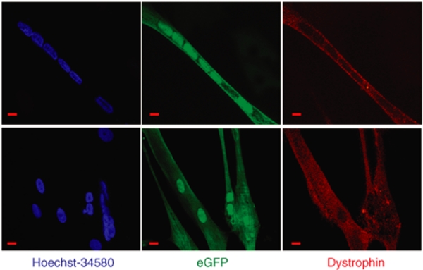
Figure 3.
hMADS and DMD cell cocultures. Dystrophin-deficient DMD myoblasts were transduced with an eGFP lentiviral vector (>90% efficiency measured 2 days after transduction) and cocultured with MyoD-hMADS cells (MOI 30). Cells were cultured on glass to allow microscopic examination and were stained at the end of the longest survival time, ~18 days under such conditions. Immunostaining of dystrophin was observed with an Alexa Fluor 594-conjugated secondary antibody. The figure shows two views of myotubes positive for both eGFP (green) and dystrophin (red) which are hybrid myotubes formed from DMD and MyoD-hMADS cells. Nuclei were labeled in blue with Hoechst-34580 and show the multinucleated structure of myotubes. Myotubes were often found isolated in the cultures because of the plating on glass, which was required for the immunostaining. Bar = 20 µm. DMD, Duchenne muscular dystrophy; eGFP, enhanced green fluorescent protein; hMADS, human multipotent adipose-derived stem; MOI, multiplicity of infection.

