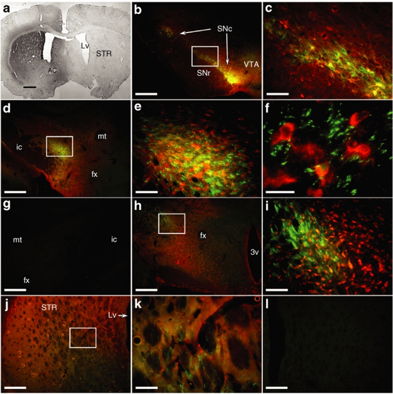
Figure 1.
GDNF distribution and viral transduction pattern. (a) Sections from an animal injected in the SNc with the CBA-GDNF/HSV-TK-GFP virus were stained for GDNF to show the level of transduction of the entire striatum using bright field microscopy, and fluorescence to show the transduction pattern with precise anatomical tracing of the tract as assessed by GFP (green) and the distribution of GDNF (red) relative to the GFP+ fibers. (b) At the level of the SN, GDNF immunoreactivity was observed throughout the SN both intra- and extracellularly, with transgene observed relatively distal to the transduced cells, arrows point to the substantia nigra pars compacta (SNc). (c) Higher magnification of area outlined in b. (d–f) GDNF immunoreactivity was observed in a wide area adjacent to the MFB throughout the nigrostriatal tract, including the posterior portions of the lateral hypothalamus. (e,f) Increased magnifications of area outlined in d. (g–i) GDNF and GFP expression at the level of the anterior hypothalamus. (i) Higher magnification of area outlined in h. Conversely, no GDNF or GFP expression was observed in the contralateral nigrostriatal tract. (j) In the terminal region of the striatum, GDNF immunoreactivity was seen throughout the striatum while a majority of GFP+ fibers were observed in the ventral portions of the striatum. (k) Higher magnification of area outlined in j. (l) Again, no GDNF or GFP expression was observed in the contralateral hemisphere. Bars in a–c = 1 mm, d, g, h, j, and l = 0.5 mm, in c, e, i, and k = 0.1 mm, and in e = 25 µm. AC, anterior commissure; fx, fornix; GDNF, glial cell line–derived neurotrophic factor; GFP, green fluorescent protein; HSV-TK, herpes simplex virus thymidine kinase; ic, internal capsule; LV, lateral ventricle; MFB, medial forebrain bundle; mt, mammillothalamic tract; STR, striatum; SNr, substantia nigra pars reticulata; VTA, ventral tegmental area; 3v, third ventricle.

