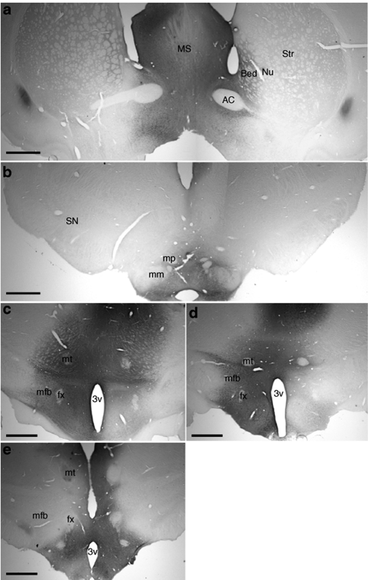
Figure 3.
GDNF expression in the hyp-GDNF group. Sections were stained for GDNF expression using a transgene specific antibody. Images were taken throughout the entire axis of transgene expression (a) stretching from the striatum, (b) to the midbrain, (c–e) throughout the hypothalamus. (a) GDNF expression in the area of the striatum at approximately the level of bregma. (b) GDNF expression at the level of the midbrain. (Bregma −4.8 mm). (c) GDNF staining at the anterior (bregma −2.8 mm), (d) medial (bregma −3.6 mm) and posterior (bregma −4.2 mm), (e) portions of the hypothalamus. AC, anterior commissure; BedNu, bed nucleus of the stria terminalis; GDNF, glial cell line–derived neurotrophic factor; MFB, medial forebrain bundle; mm, medial mammilary nucleus; mp, mammillary peduncle; MS, medial septum; Str, striatum, SN, substantia nigra; Bar = 1 mm.

