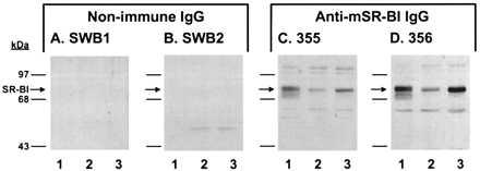
Figure 1.

Western blot analysis of anti-mSR-BI antibodies. Postnuclear supernatant (20 μg protein) from ldlA[mSR-BI] cells (lane 1), and Y1-BS1 cells treated without (lane 2) or with (lane 3) 1–24ACTH were separated by SDS/8% PAGE and transferred to nitrocellulose membranes. The membranes were incubated overnight at 4°C in the presence of either SWB1 nonimmune IgG (A), SWB2 nonimmune IgG (B), 355 anti-mSR-BI IgG (C), or 356 anti-mSR-BI IgG (D) at 4 μg/ml. IgG binding was visualized by enhanced chemiluminescence.
