Figure 3.
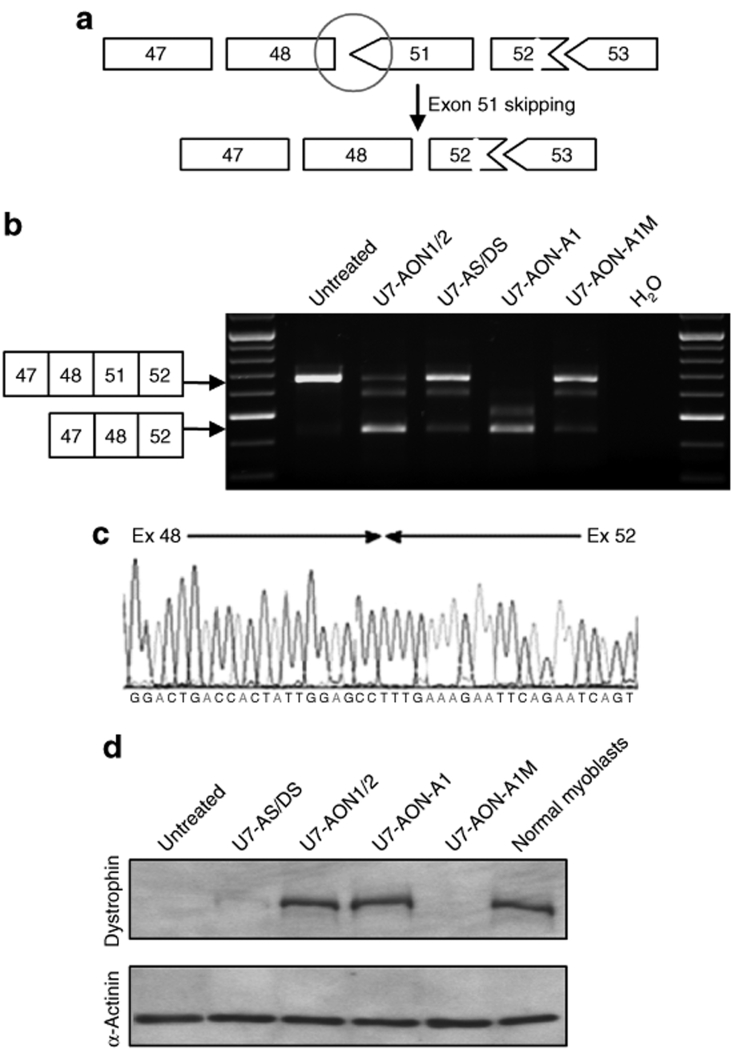
Comparison of exon 51 skipping in DMD myoblasts and subsequent dystrophin rescue. (a) Schematic representation of the Δ49–50 DMD deletion, which creates a codon de-phasing between the exons 48 and 51 (black circle) and the expected link generated by exon 51 skipping. (b) DMD myoblasts carrying the Δ49–50 deletion were transduced with lentiviral vectors encoding the different U7 snRNA constructs. Exon 51 skipping was assessed by nested reverse transcriptase–PCR and a fragment of the expected size was detected in each case. Additional bands due to heteroduplex formation and activation of a cryptic splice site within the exon 51 (previously described34) are visible in same analyses. (c) Bands were analyzed by sequencing confirming the precise skipping of exon 51, joining exons 48 and 52. (d) Western blot probed with dystrophin antibody (top gel) and α-actinin antibody (bottom gel). Note that the dystrophin detected corresponds to an internally deleted protein missing exons 49, 50, and 51. AON, antisense oligonucleotide; AS, acceptor splice; DMD, Duchenne muscular dystrophy; DS, donor splice.

