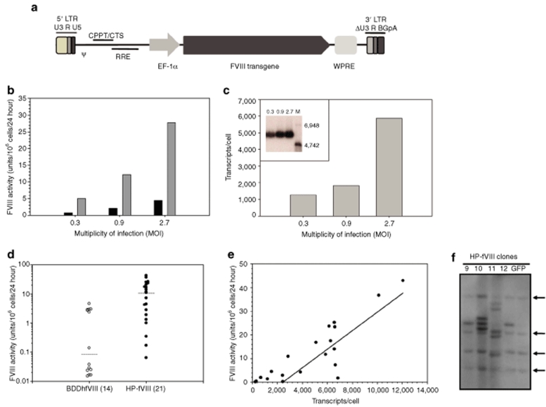
Figure 2.
FVIII expression analysis following lentiviral transduction of HEK-293T cells. (a) HIV-1-based lentiviral vectors were manufactured by Lentigen using the LentiMax Production System. LTR, long terminal repeat; U3, R and U5, promoter/enhancer regions; psi, packaging signal; cPPT, central polypurine tract; RRE, rev response element; eF1-α, elongation factor 1-α promoter; WPRE, woodchuck hepatitis post-transcriptional regulatory element; ΔU3, modified LTR for production of recombinant self-inactivating lentiviral vectors. (b) FVIII expression levels were determined from transduced HEK-293T cells expressing HP-fVIII (shaded bars) or BDDhfVIII (black bars), which was determined by a one-stage clotting assay. Each bar represents the mean activity of two wells. (c) Average transcripts per cell were measured by quantitative PCR from HP-fVIII transduced HEK-293T cells at MOI 0.3, 0.9, and 2.7 (shaded blocks), and northern blot (c, inset) analysis of the same cells indicating fVIII transcripts of approximately 6,300 nucleotides. (M denotes molecular weight marker with sizes as indicated.) (d) HP-fVIII and BDDhfVIII HEK-293T expressing clones were generated by limiting-dilution cloning and fVIII activity levels were measured by one-stage coagulation assay (P < 0.001). Horizontal lines indicate the mean values for each group and the number in parenthesis indicates the number of clones analyzed. (e) The number of HP-fVIII transcripts per cell were determined by quantitative RT-PCR and plotted against fVIII activity from a one-stage clotting assay demonstrating a correlation between transcript number and fVIII activity (r2 = 0.765, P < 0.0001). (f) Southern blot analysis was performed on genomic DNA isolated from clones generated in d and digested with AvrII. The arrows indicate endogenous fVIII DNA fragments.

