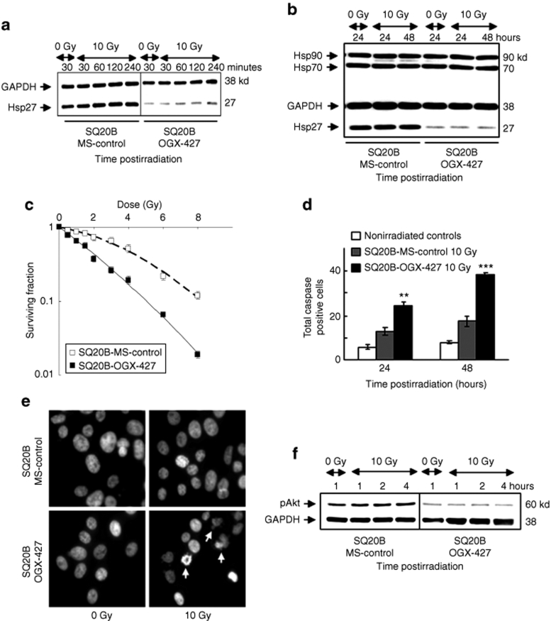
Figure 1.
Knockdown of Hsp27 by OGX-427 sensitizes SQ20B cells to irradiation. (a) Immunoblot analysis of Hsp27 expression in response to 0 or 10 Gy irradiation of SQ20B-MS-control and SQ20B–OGX-427 cells. (b) Immunoblot analysis of Hsp27, Hsp70, and Hsp90 protein expression in response to 0 or 10 Gy irradiation. Protein (10 µg) was loaded on the gels. Glyceraldehyde-3-phosphate-deshydrogenase (GAPDH), as loading control. (c) Cell survival after exposure of SQ20B-MS-control and SQ20B–OGX-427 cell lines to radiation at doses varying between 0 and 8 Gy. Colonies with >64 cells were scored after six cell divisions following irradiation. (d) Total caspase activity quantified by flow cytometry. Each value represents the mean ± SD of two experiments performed in triplicate. **P < 0.005, ***P < 0.001 versus irradiated SQ20B-MS-control cells. Nonirradiated controls represent the mean of results (triplicate) obtained in basal conditions for each transfected cell line. (e) Fluorescent DAPI staining examined 48 hours postirradiation. Apoptotic cells appear with characteristic disintegrated chromatin in the nuclei (white arrows) (original magnification ×40). (f) Immunoblot analysis of the phosphorylated active form of Akt (pAkt) in response to 0 or 10 Gy irradiation of SQ20B-MS-control and SQ20B–OGX-427 cells. DAPI, 4′-6-diamidino-2-phenylindole-dihydrochloride; GAPDH, glyceraldehyde-3-phosphate-dehydrogenase; Hsp, heat shock protein.

