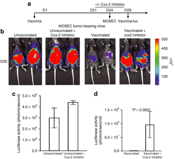
Figure 2.
Luminescence imaging of MOSEC tumor-bearing mice infected with luciferase-expressing vaccinia virus with or without treatment of Cox-2 inhibitor. (a) Schematic diagram demonstrating the regimen of vac-luc and Cox-2 inhibitor treatment in MOSEC tumor-bearing mice previously infected with wild-type vaccinia. C57BL/6 mice (five per group) were infected with wild-type vaccinia at a dose of 1 × 10⁷ pfu/mouse on day 1 (D1). Mice were treated with or without Cox-2 inhibitor (100 mg/kg per day) orally from D21 to D28. Mice without the first vaccination treated with or without Cox-2 inhibitor served as controls. All groups of mice were then challenged with 2 × 10⁶ MOSEC cells/mouse on D24 and infected with vac-luc at a dose of 1 × 10⁷ pfu/mouse on D28. Mice were imaged using the IVIS Imaging System Series 200. (b) Representative bioluminescence images of MOSEC tumor-bearing mice treated with vac-luc ± Cox-2 inhibitor. Images were acquired on D30. (c) Bar graph illustrating the luciferase activity (photons/second) in MOSEC tumor-bearing mice treated with or without Cox-2 inhibitor. (d) Bar graph illustrating the luciferase activity (photons/second) in MOSEC tumor-bearing mice treated with vac-luc ± Cox-2 inhibitor (*P < 0.0001). Data shown are representative of two experiments performed (mean ± s.d.).

