Fig. 3.
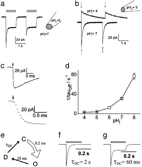
Activation of ChR2 photocurrents in excised inside-out giant membrane patches from Xenopus oocytes. This configuration allows a rapid concentration change (≈100 ms; ref. 43) at the cytoplasmic side by changing the superfusing bath solution. (a) Activation of inward photocurrents by light pulses of 442 nm (gray bars). Pipette solution used was 115 mM NMG-Cl, pH 5; bath (cytoplasmic solution) used was 115 mM NMG-Cl, pH 7. Currents typical of those in nine other experiments are shown. (b) Activation of photocurrents by two laser flashes of 10-ns duration (arrows), 440 nm. Pipette solution used was 115 mM NMG-Cl, pH 5; bath solution used was 115 mM NMG-Cl, pHi 7or 115 mM NMG-Cl, pHi 4, as indicated. Currents typical of those in three other experiments are shown. (c) Higher time resolution of first photocurrent, shown in b, pHi = 7. (d) Dependence of closing rate on pHi (n = 4–7). (e) Simple three-state model of ChR2 photocycle. Light, curved arrow indicates light-activated step from ChR2-ground state (C) to open state (O). Straight arrows indicate dark reactions, to the closed desensitized state (D) and back to the ground state (C). For further details see text. (f) Simulated photocurrent with τDC = 2 s and other time constants as in the model depicted in e. (g) Simulated photocurrent with τDC = 60 ms and other time constants as in e.

