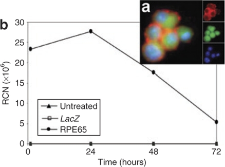
Figure 1.
Hematopoietic stem cells (HSCs) infected with RPE65-lentivirus construct for 2 hours resulted in expression of RPE65 mRNA and protein. (a) Immunofluoresence micrograph shows RPE65 protein expression by infected HSCs. The large image is a merge of the separate channels shown in the insets. Blue is nuclear staining from 4′,6-diamidino-2-phenylindole, green is native green fluorescent protein, and red is RPE65 detected by anti-RPE65 antibody followed by rhodamine-conjugated second antibody. (b) Time course of mRNA expression for HSCs infected with RPE65 lentivirus. mRNA was harvested from the infected cells either immediately after the 2-hour infection period ended, 1, 24, 48, and 72 hours after the end of the 2-hour infection period. The mRNA was reverse transcribed using WT-Pico Kit (NuGEN, San Carlos, CA) and amplified using real-time PCR with the human gene–specific primers for RPE65 (TaqMan; ABI). Data were normalized to GSTp1 (detected by GeneChip as having a very low coefficient of variation throughout the experiment) and expressed as relative copy number (RCN). Note that even after 72 hours, the level of expression remained in the 5 × 10⁶ range. For the same time points, the mouse RPE65 was not detectable by TaqMan assay (data not shown).

