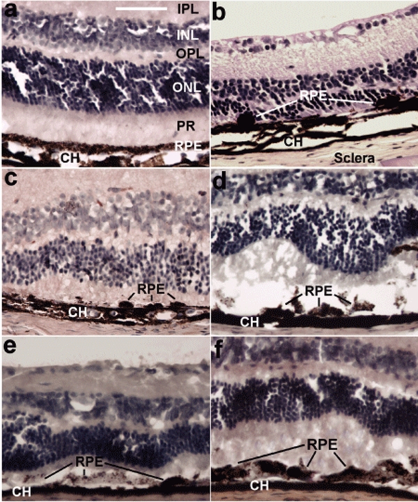
Figure 3.
Hematoxylin and eosin–stained cross-sections showed gross damage from sodium iodate and apparent rescue of retinal pigment epithelium (RPE) and neural retina anatomy by RPE65-transfected hematopoietic stem cells (HSCs). All images are of paraffin-embedded whole globes, cut at 10 µm thickness. (a) A normal uninjured eye with integrity of the RPE layer and photoreceptors. (b) An eye that was injected with 100 mg/kg sodium iodate but was not given any rescuing HSCs. Note the complete absence of the photoreceptor outer segments, near absence of any RPE cells (indicated by lines), and profound decrease in the retinal thickness. (c–f) Cross-sections of eyes from animals that were all given the same dose (100 mg/kg) of sodium iodate, and were also given (c) uninfected HSCs, (d) LacZ-infected HSCs, or (e,f) RPE65-infected HSCs. Animals receiving uninfected HSCs demonstrated only occasional RPE cells on Bruch's membrane at 28 days (c). Animals receiving LacZ-infected HSCs showed a small increase in cells compared to uninfected HSCs but large patches of Bruch's membrane were devoid of cells even at 28 days (d). By contrast, animals receiving RPE65-infected HSCs demonstrated abundant cells on Bruch's membrane. Seven days after treatment, a disorganized and poorly pigmented monolayer of cells was observed (e) and by 28 days (e), a mature highly pigmented epithelial layer had formed on Bruch's membrane. This layer exhibited all the characteristics of native RPE; photoreceptor outer and inner segments were also apparent. Bar = 70 µm. CH, choroid; INL, inner nuclear layer; IPL, inner plexiform layer; ONL, outer nuclear layer; OPL, outer plexiform layer; PR, photoreceptor inner and outer segments.

