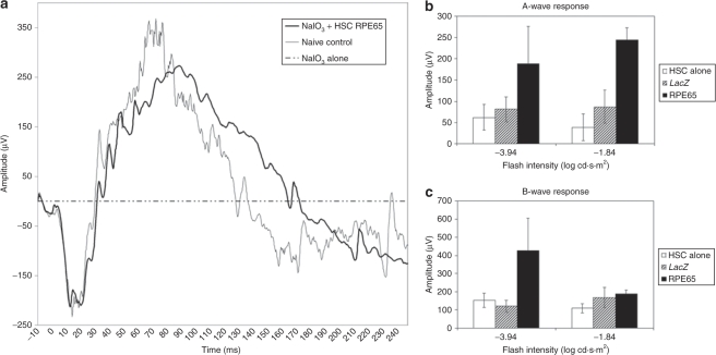
Figure 7.
Electroretinography (ERG) response demonstrated the RPE65-transfected HSC-driven rescue of visual function after sodium iodate damage. (a) Typical ERG traces from untreated (naive) control mice and from sodium iodate–injured animals 28 days after receiving HSCs expressing hRPE65. Sodium iodate alone causes a complete loss of normal ERG response. In marked contrast eyes from animals that received HSCs expressing hRPE65 have retinal function that is almost identical to the naive control, in both the A-and the B-wave amplitude. Each of the traces represents the averaged signal from five flashes at a light intensity of −1.84 log cd·s·m2. (b,c) The mean A-wave and B-wave amplitudes, respectively, for low and high flash intensities averaged for n ≥ 5 mice per treatment condition. Animals receiving HSCs expressing hRPE65 after sodium iodate injury show a statistically significant improvement in both A- and B-wave response compared to animals receiving either uninfected HSCs or HSCs expressing LacZ. *P < 0.05.

