Figure 4.
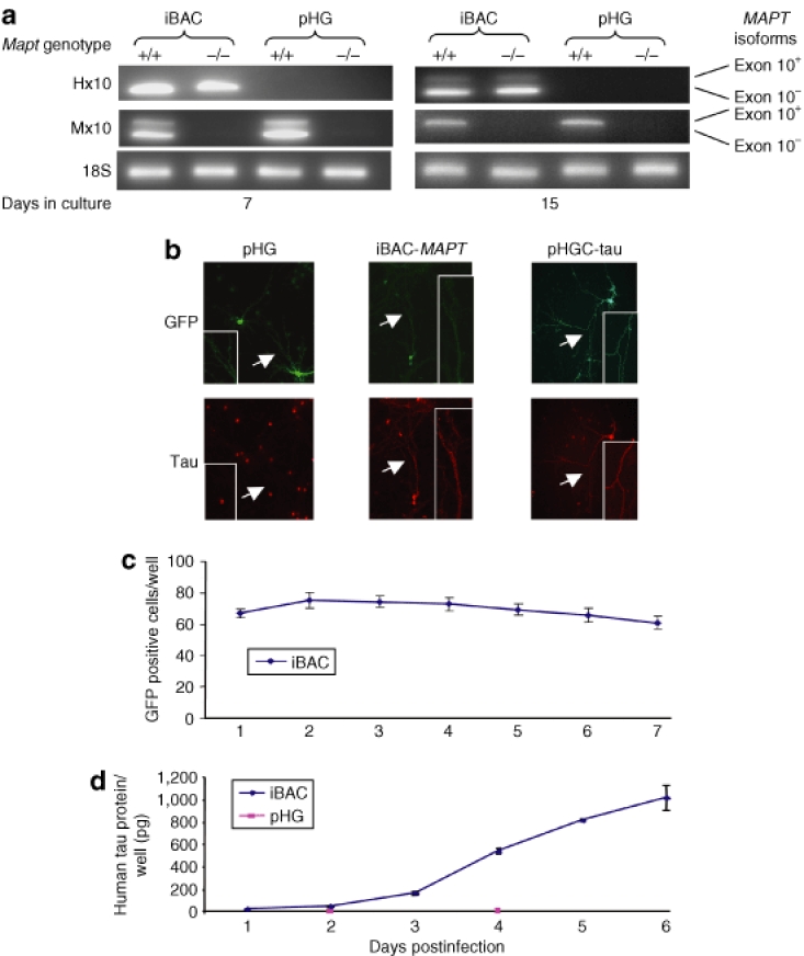
iBAC-MAPT transgene RNA and protein expression in mouse Mapt−/− primary neurons. (a) The iBAC-MAPT transgene RNA is expressed in primary neurons prepared from Mapt−/− P0 pups. P0 mouse primary cultures were transduced with iBAC-MAPT (MOI = 1) after either 5 or 13 days in culture. RNA was extracted 2 days post-transduction and species-specific RT-PCR was performed. The human exon 10+(4R) MAPT form is expressed in a developmentally regulated way, only present in mature neurons after 15 days in vitro. Primer pairs: Hx10, human exon 10; Mx10, mouse exon 10. (b) Immunocytochemistry confirms expression of tau protein in Mapt−/− neurons following transduction by iBAC-MAPT, but not pHG. P0 mouse Mapt−/− primary cultures were transduced with iBAC-MAPT or control vectors (MOI = 1) after 5 days in culture and immunocytochemistry was performed 9-days post-transduction. pHGC-tau is a positive control amplicon vector expressing human MAPT complementary DNA. Axonal staining of GFP and tau protein is indicated by an arrow and is seen in detail in the inset. The red cell-body staining is a nonspecific signal associated with the secondary antibody and seen in Mapt−/− neurons transduced with the pHG negative control vector. (c) Counts of GFP positive cells over time following iBAC-MAPT transduction. Prolonged iBAC vector backbone-encoded GFP expression confirms prolonged vector retention. (d) Tau protein levels per well measured by enzyme-linked immunosorbent assay over time following iBAC-MAPT or pHG transduction. iBAC-MAPT maintains prolonged tau protein expression leading to a steady increase in tau protein levels in tranduced cells. (c) and (d) P0 mouse Mapt−/− primary cultures were transduced after 5 days in culture and transduced at MOI = 1.

