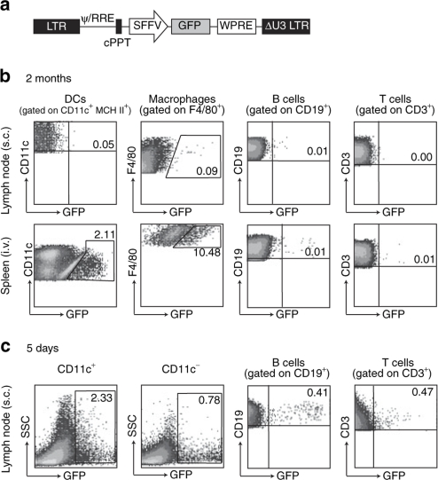
Figure 1.
Biodistribution of transduced cells. (a) Mice (2–3 per group) were injected either subcutaneously (s.c.) or intravenously (i.v.) with phosphate buffered saline or 108 IU GFP LV. At different time points, cells from spleen and lymph nodes (inguinal and popliteal) were collected, pooled, separated with anti-CD11c magnetic microbeads and stained with antibodies for flow cytometric analysis. The percentage of GFP+ DCs, macrophages, B cells and T cells was determined in (b) the lymph nodes and spleen 2 months after s.c. or i.v. injection, and (c) in lymph nodes 5 days after s.c. injection. DC, dendritic cell; GFP, green fluorescence protein; LV, lentiviral vector.

