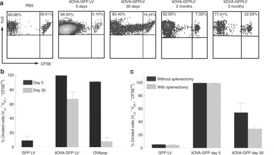
Figure 4.
Persistence of antigen presentation. Mice received intravenous injection of 5 × 10⁶ CFSE-labeled cells from OT-1 mice at different time points after intravenous injection of PBS, GFP or IiOVA-GFP LVs. Five days later, transferred cells were tracked in the spleen and their expansion was assessed by dilution of CFSE by flow cytometry. The plots show events gated in (a) Vα2+ Vβ5.1+ cells. (b) Results from five experiments are summarized. (c) The experiment was repeated but 2 days before the adoptive transfer, spleens were removed and the OT-1 cells were tracked in peripheral blood. CFSE, carboxyfluorescein succinimydil ester; GFP, green fluorescence protein; LV, lentiviral vector; OVApep, OVA peptide; PBS, phosphate buffered saline.

