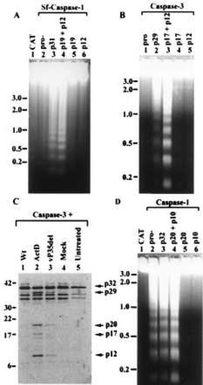
Figure 1.

Induction of apoptosis and caspase processing activity in Sf-21 cells. (A, B, D) DNA isolated from Sf-21 cells 10 h after transfection with plasmids expressing various caspases and their forms, as indicated in the figure, was electrophoresed through a 1.2% (A) or 1.5% (B, D) agarose gel and visualized by ethidium bromide staining. CAT-transfected cells (lane 1 of A and D) served as negative control for induction of apoptosis. (C) Purified, in vitro translated, and 35S-labeled pro-caspase-3 was incubated for 12 h with cell extracts prepared from Sf-21 cells treated as follows: wt baculovirus-infected (Wt, lane 1), actinomycin D-treated cells (Act D, lane 2), vP35del-infected (lane 3), mock-infected cells (lane 4). Untreated, purified in vitro-translated pro-caspase-3 is shown in lane 5. Samples were resolved on a SDS/12% polyacrylamide gel and visualized using a PhosphorImager. Size standards are indicated to the left of the gel. Arrows on the right show the position of pro-caspase-3 and known processed forms of caspase-3. Extra bands in the untreated lanes are other products of the in vitro translation reaction including products possibly initiated from internal methionine codons.
