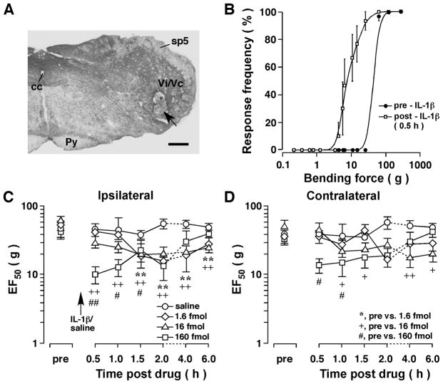
Figure 1.
Mechanical hyperalgesia/allodynia induced by injection of IL-1β into the Vi/Vc transition zone of the rat. A. An example of caudal brain stem section stained with cresyl violet for histological verification of the site of microinjection. Arrow indicates the injection site in ventral Vi/Vc. Scale bar = 500 μm. cc, central canal; Py, pyramidal tract; sp5, spinal trigeminal tract; Vi/Vc, trigeminal subnuclei interpolaris/caudalis transition zone. B. Stimulus-response function curves illustrating the intensity-dependent head withdrawal responses to mechanical stimuli. Each curve was established with a series of subthreshold to suprathreshold range of von Frey filament forces and the response frequency is plotted against the stimulus intensity. IL-1β (160 fmol) was injected into the ventral Vi/Vc. The skin site above the masseter muscle was probed. Note that there was a leftward shift of the curve at 30 min after IL-1β injection compared to the pre-IL-1β curve (p<0.01), suggesting the development of mechanical hyperalgesia and allodynia. Best-fit curves were generated by nonlinear regression analysis (GraphPad Prism). C, D. The EF50s were derived from the respective stimulus-response frequency function curves and are plotted against time. Note significant decreases in EF50s at 30 min-6h after IL-1β injection, indicating IL-1β̃ induced bilateral hyperalgesia/allodynia. +, #: p<0.05; **, ++, ##: p<0.01 (ANOVA with repeated measures and post-hoc test). Dashed lines indicate interruption of the linearity of the time scale.

