
Figure 3. Pluripotent capacity and embryonic developmental potential in 4F-iPS and 3F-iPS.
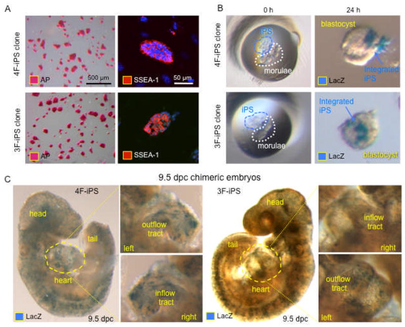
A, 4F-iPS and 3F-iPS stain with similar positive patterns for pluripotency markers alkaline phosphatase (left, AP) and SSEA-1 (right, nuclei revealed with DAPI). B, LacZ-labeled iPS coincubated with diploid embryos (left) revealed the ability of 4F-iPS and 3F-iPS to integrate into host blastocyst (right). C, Integration of both 4F-iPS and 3F-iPS was sustained through embryonic development as shown for 9.5 dpc embryos, containing labeled cells that contributed to most of the tissues including the heart (cardiac inflow and outflow tracts showed in insets).