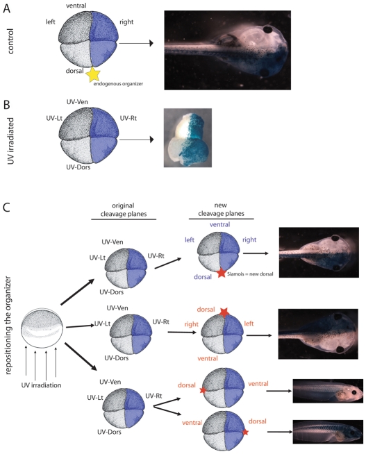
Fig. 2.
Establishment of the midline is dependent on the location of the new organizer, unless no organizer is present. (A) Both right blastomeres of control four-cell embryos were injected with β-gal. When the organizer develops on the dorsal side as expected (termed the ‘endogenous placement’, marked by a yellow star), β-gal signal remains localized to the right side of the tadpole. (B) In UV-irradiated embryos, the blastomeres no longer have ‘dorsal’ or ‘ventral’ fates, although they maintain the same appearance (i.e. cell size and pigmentation). New terms are therefore assigned to each blastomere (i.e. UV-Ven, UV-Dors, UV-Rt, UV-Lt). β-gal was injected into both UV-Rt blastomeres at the four-cell stage. If no further treatment is given, the β-gal signal localizes to one half of the resulting belly piece. (C) Both UV-Rt blastomeres of four-cell UV-irradiated embryos were injected with β-gal. Then, the position of the organizer was specifically targeted to one of three locations (marked by a red star) via injection of XSiamois at the 16-cell stage. When XSiamois is injected into the same location as the endogenous placement (UV-Dors), the cells maintain their original identities and the β-gal localizes to the right side of the rescued tadpole. When XSiamois is injected opposite the endogenous placement, the UV-Ven cells are re-defined as dorsal, and the β-gal signal localizes to the left side of rescued tadpole. Finally, if the organizer is positioned 90 degrees from the endogenous placement, β-gal is localized to either the ventral or the dorsal cells. Thus, proper organizer placement could be verified by β-gal localization in rescued tadpoles. Labels for cells that maintain their original identity are shown in blue, labels for cells that changed identity after placement of the organizer are shown in red.

