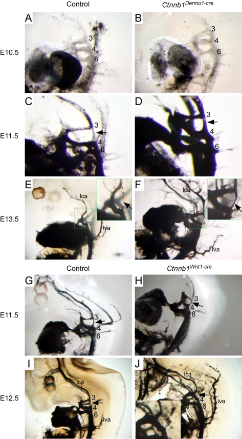
Fig. 3.
Mesenchymal β-catenin is required for normal PA artery remodeling. (A,B) At E10.5, both control (A) and Ctnnb1Dermo1-Cre (B) embryos showed similar aortic arch artery structures. (C,D) At E11.5, there is a normal atrophy of the dorsal aorta between PA artery 3 and 4 (C, arrow). Ctnnb1Dermo1-Cre mutant embryos (D) do not show regression of the PA artery 3 and 4 channel (arrow). (E,F) By E13.5, the dorsal aorta between PA artery 3 and 4 is normally lost (E, red dashed line, arrow in inset). Ctnnb1Dermo1-Cre mutant embryos (F) retained the dorsal aorta between PA artery 3 and 4 (arrow in inset). (G-J) In Ctnnb1Wnt1-Cre mutant embryos (H,J), the PA artery remodeling occurred normally with no apparent difference from controls (G,I) at E11.5 and E12.5, respectively. Arrows indicate the dorsal aorta spanning PA artery 3 to 4. 3, third pharyngeal arch; 4, fourth pharyngeal arch; 6, sixth pharyngeal arch; ia, innominate artery; lca, left common carotid artery; lva, left vertebral artery; rca, right common carotid artery.

