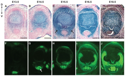
Fig. 7.
Formation of the metacarpophalangeal joint and development of the skeletal and tendon components within it. (A-E) Skeletal precursor cells labeled by X-Gal staining on MP joint sections for Sox9Cre/+; R26R/+ embryos at the stages indicated. (F-J) Tendon/ligament progenitors are visualized by green fluorescence on MP joint sections from Scx-GFP/+ transgenic embryos. At E13.5, skeletal and tendon cells are largely symmetrically distributed along dorsal-ventral axis. The dorsal-ventral asymmetry starts to appear because of the presence of the FDS/FDP tendon (G, arrow) from E14.5 and of the ventral sesamoid bone (C, arrows) at E15.5. The whole MP structure is fully developed by E18.5. Scale bars: 100 μm.

